The Biologics of Degenerative Disc Disease, Part III

In our third and final post of this series we’ll discuss some of the difficulties in using mesenchymal stem cells (MSCs) for regenerative therapies for degenerative disc disease (DDD). To recap from the last post, here’s what we know so far about MSCs in the treatment of DDD: First, we know that MSCs can be safely and efficiently harvested and cultured to a quantity of cells suitable for implantation.  We also know that these MSCs can be cultured in vitro (in a test tube) into cells similar to nucleus pulposus (NP) cells.  We know that MSCs can be safely implanted into an intervertebral disc (IVD) of animals or humans.  Finally, we know that implantation of MSCs can lead to improvement in the cellular appearance and MRI appearance of the IVD.  In some very preliminary human studies this has translated into some clinical benefit.  Sounds like we’ve got this all worked out right?  
 
Unfortunately, we’re not quite there yet.  It’s long been know that the problem with stem cells isn’t getting them to a location, it’s getting them to stay there and flourish.  Stem cell scientists will frequently report that when they survey a site of implantation weeks after MSCs are implanted there are few cells left.  We suspect that this is because once the cells are injected, if they don’t have the proper nutritional and structural support they will undergo apoptosis (cell suicide) and die. The next step in getting these regenerative therapies to work, then, is to ensure that the injected MSCs have what they need to survive and produce the extracellular matrix (ECM) necessary for IVD regeneration.
 
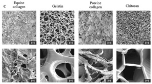
First, it’s clear from many years of in vitro stem cell work that MSCs prefer three-dimensional organization to thrive.  Stem cell scientists often culture MSCs with beads of aliginate, a non-soluble gelatinous substance that acts a scaffold to promote proliferation.  Recently more complex scaffolds have been developed.  These scaffolds are composed of materials ranging from simple, naturally-derived collagen to exotic nano-designed proprietary materials (see figure 1).  The materials not only have to be able to provide a hospitable locale for the MSCs, but also have to be structurally robust so that they can stand up to the mechanical stresses in the IVD.  Ultimately, though, the goal of these various materials is the same: to provide structural support to anchor the MSCs so that they can differentiate into cells necessary for IVD regeneration.  Future therapies will likely include some sort of scaffold material injected concurrently with the MSCs.
 
Mendeley Desktop
Figure 1. Various types of scaffolds used for MSC culture.  (Source: Bertolo et al, 2012)
 
Another obstacle to in vivo (in the body) MSC differentiation and proliferation is that the environment within the IVD is quite harsh.  Even in a young healthy IVD the blood supply that provides oxygen and nutrients to the disc is quite tenuous.  Over time with repeated mechanical loading it is believed that this delicate blood supply (most of which comes through the bony endplates of the adjacent vertebral bodies) is damaged resulting in the death of IVD cells.  Some groups have experimented with various substances (i.e. calcium channel blockers) to help increase blood flow to the disc.  Others have looked at mechanically altering the adjacent endplates to allow more blood to flow to the IVD.  We have quite a way to go before any of these therapies are ready for clinical use, however.
 
Ok let’s say that you’ve gotten the MSCs to stay in place on a nice scaffold.  You’ve also figured out a way to augment blood flow and nutrition to the disc space.  How do you actually get the MSCs to differentiate into the various cell types of the IVD?  Native NP cells produce ECM that has a very specific ratio of proteoglycans (the water-carrying substance so important to healthy IVDs) and collagen (27:1 for those interested.)  Unfortunately, using current culture methods MSCs often will by default differentiate into cells similar to chondrocytes, cells that form cartilage.  This results in the formation of ECM that is thick and fibrous like the cartilage in a knee rather than the soft and pliable ECM of an NP.  Researches are getting closer, however, to the proper culture techniques that promote the differentiation of MSCs into cells nearly identical to NP cells.  Some of these techniques involve the implantation of a concoction of MSCs and specific growth factors that promote proper differentiation.  Other techniques utilize special scaffolds that secrete growth factors to stimulate attached MSCs.  These techniques have shown promise in animal studies.  One last thing to consider: remember that the NP is only one component of the IVD and that the annulus fibrosus (AF) is equally important for the structure and function of the IVD.  Despite this fact, almost no one is working on the differentiation of the MSCs into AF-like cells. We clearly have a long way to go before we can reliably direct implanted MSCs to differentiate into cells of each distinct component of the IVD.     
 
Perhaps rather than trying to coax foreign cells to become NP cells we should be trying to find cells within the IVD that have the potential to form the various IVD cell types.  Every tissue type in your body is composed of cells that originate from parent progenitor cells.  These progenitor cells are originally quite pluripotent in that they are able to differentiate into a variety of related cell types.  While most of these progenitor cells die off as we mature, a small number remain dormant within the tissue in case they’re needed for purposes of regeneration.  It turns out that the IVD also has nests progenitor cells.  Recently a group in Japan (Sakai et al, 2012) discovered a population of cells, so-called Tie+ cells, that can differentiate into mature NP cells.  The fact that the survival of these cells was dependent on angiopoietin-1, a potent promoter of blood vessel formation, stresses the importance of adequate blood flow on the health of the cells within the IVD.  These progenitor cells may be the key to unlocking the mystery of IVD regeneration.
 
Readers of this post will realize that many hurdles still must be overcome in the development of stem cell therapies for IVD regeneration. Regardless, this is a very exciting frontier in the treatment of DDD.  A review of any current spine journal reveals a new in vivo or animal study of these therapies.  Also, a search of clinicaltrials.gov lists several clinical trials enrolling patients to study some sort of implantable stem cell therapy for DDD.  Clearly momentum is building and it won’t be long before stem cell therapies for the treatment of DDD become reality.  
 
Thanks for reading!
 
J. Alex Thomas, M.D.
 
1. Sakai D, Nakamura Y, Nakai T, Mishima T, Kato S, Grad S, et al.: Exhaustion of nucleus pulposus  progenitor cells with ageing and degeneration of the intervertebral disc. Nat Commun 3:1264, 2012. 

2. Bertolo A, Mehr M, Aebli N, Baur M, Ferguson SJ, Stoyanov J V: Influence of different commercial scaffolds on the in vitro differentiation of human mesenchymal stem cells to nucleus pulposus-like cells. Eur Spine J 21 Suppl 6:S826–38, 2012.
The Biologics of Degenerative Disc Disease, Part II: Stem Cell Therapies

Perhaps nothing captures the imagination of scientists and the general public alike more than the potential of stem cell therapies.  These therapies direct pluripotent cells (i.e. cells that can become any cell in the body) to replenish cells that have been damaged or depleted by disease processes.  Think diabetes, Parkinson’s Disease, ALS and even spinal cord injury.  While the reality of stem cell therapies hasn’t always lived up to our expectations, the field of stem cell therapy research is still in its infancy and I think we have only begun to see the power of these therapies.  One of the areas where stem cell therapies are starting to gain traction is in the treatment of DDD.  

In our last post we discussed how a concoction of growth factors and cytokines maintains the health of the structural components of the intervertebral discs (IVDs).  (For simplicity’s sake we’ll refer to these structural components wholly as extracellular matrix, or ECM.)  These growth factors and cytokines stimulate cells within the IVD to produce ECM.  For reasons that are not yet fully understood the populations of these cells, and thus amounts of ECM they produce, are depleted over time.  This is where we get a “Chicken or Egg” scenario: is degenerative disc disease (DDD) caused by the loss of these cells or is some other trigger of DDD (i.e. years of mechanical stress) the cause of the loss of these cells.  I’m not sure that the answer to this question is know yet, but what is known is that these populations of cells are tremendously important to the health of the IVD. The goal of stem cell therapies for DDD is to replace or regenerate these populations of IVD cells with the goal of replenishing the ECM components that give the disc its form and function.  Sounds simple, right? 

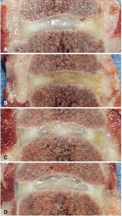
It turns out that it’s actually quite complicated.   The first obstacle to overcome is where to get the “starter” cells used to replenish the lost populations of IVD cells.  Probably the most straightforward way to replenish these cells is to just find a source of mature IVD cells for harvest.  Some studies have looked at harvesting cells from herniated pieces of disc material removed at surgery.  The number of cells within the IVD is quite low (they make up only 1% of the total disc volume) so after the cells are harvested from the disc fragments they must be cultured and multiplied until millions of cells are present. The cells can then be transplanted back into the diseased disc.  Animal studies have confirmed that this is a viable method of increasing the number of IVD cells present and that these cells can improve the health of the IVD (See Figure 1).  In one human study, the EuroDisc trial, cells were cultured and multiplied to over 5 million cells and then transplanted back into diseased discs at 12-weeks (so another procedure was required.)  Although no placebo was used and there certainly were flaws in the assessment of outcomes, an initial analysis of EuroDisc data in 2008 revealed a decrease in pain and increase in disc hydration (as measured with MRI) at 2-year follow-up in patients who underwent cell transplantation versus controls.  A more thorough analysis of their data was due out a few years ago but to my knowledge hasn’t been released.  
Mendeley Desktop

Figure 1. Specimens from dogs treated during animal phase of EuroDisc investigations.  These are gross specimens analyzed 6-months after treatment.  Image A is a control injured disc, note the dusky color, height loss and loss of distinction of the central nucleus pulposus. Image B is an injured disc injected with hyaluronic acid, looks even worse. Image C is an injured disc injected with adipose-derived stem cells while image D is a normal, uninjured disc.  Note how the color, height and nucleus pulposus are preserved. (Source: Hohaus C, Ganey TM, Minkus Y, Meisel HJ: Cell transplantation in lumbar spine disc degeneration disease. Eur Spine J 17 Suppl 4:492–503, 2008. Figure 6.)

Another way to grow a population of IVD cells is to start with true, pluripotent stem cells.  These cells, capable of growing into a variety of tissue types, can be directed to become mature IVD cells. One main benefit of using stem cells as starter cells is that they can be easily obtained from the patient without the need for a spinal procedure to remove a piece of herniated disc to harvest cells from.  The stem cells we’re discussing here aren’t embryonal stem cells which are harvested from an aborted fetus and thus are morally objectionable to some.  Rather, these stem cells are mesenchymal stem cells (MSCs).  The standard site of harvest of these MSCs is the bone marrow although other sites such as adipose tissue are becoming popular as well (so as a bonus you can get a liposuction during cell harvest!).  Once MSCs are harvested they are cultured under specific conditions to become cells similar to those native to the IVD.  These cells are then reimplanted into diseased discs to, it is hoped, regenerate the structural integrity of the disc.  The feasibility of these methods has been confirmed in several animal studies.  One pilot study done in humans (Orozco et al, 2011) has demonstrated not only feasibility and safety but also favorable clinical results in 10 patients with chronic back pain implanted with harvested MSCs.  

In our third and final post of this series we’ll discuss some of the difficulties with MSC transplantation as well as some novel strategies that help overcome these difficulties. 

Thanks for reading!

J. Alex Thomas, M.D.

Sources:

1. Chan SCW, Gantenbein-Ritter B: Intervertebral disc regeneration or repair with biomaterials and stem cell therapy–feasible or fiction? Swiss Med Wkly 142:w13598, 2012 Available: http://www.ncbi.nlm.nih.gov/pubmed/22653467. Accessed 17 July 2014

2. Hohaus C, Ganey TM, Minkus Y, Meisel HJ: Cell transplantation in lumbar spine disc degeneration disease. Eur Spine J 17 Suppl 4:492–503, 2008.

3. Orozco L, Soler R, Morera C, Alberca M, Sánchez A, García-Sancho J: Intervertebral disc repair by autologous mesenchymal bone marrow cells: a pilot study. Transplantation 92:822–8, 2011.

4. Sivakamasundari V, Lufkin T: Stemming the Degeneration: IVD Stem Cells and Stem Cell Regenerative Therapy for Degenerative Disc Disease. Adv Stem Cells:2013.